
Chest X-ray Interpretation
A Clinicians Perspective
Jeff Wilson MD
OVERVIEW
Goal – to show how you can read chest x-ray
Technique – brief, for clinicians
Interpretation system – You should have one
Beyond the Chest X-ray
Lessons learned
Some highlights:
-
Airways you can are the trachea and the main-stem bronchus. Left main-stem comes out at sharper angle than the right. This is why people aspirate to the right lower lung and ET tubes are often placed in the right main-stem bronchus if advanced too far.
-
Tracheal deviation away from pathology indicates positive pleural pressure shifting the trachea contralaterally. This may occur with large pleural effusion or tension pneumothorax
-
Tracheal deviation towards pathology indicates negative pleural pressure that deviates the trachea towards pathology. This may occure from collapse, lug resection, or scarring of the lung or pleura.
-
On inspiratory film, you should be able to count 9-10 ribs
-
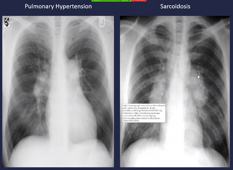
Smoothness or contour of the enlarge mediastinum may point you towards various pathology. See below the smooth contour in pulmonary hypertension in contrast to the irregular borders seen in sarcoidosis.
-
Lateral films can give lots of added information and should be obtained when able. The mediastinum can be broken down into three compartments: anterior, middle, and posterior.

-
Diaphragm is usually 1-2 cm higher on the right side.
-
Upright chest films are sensitive to note free air under the diaphragm; seen in perforated viscus, abdominal surgery, peritoneal dialysis. This is still not a perfect test with roughly 70% sensitivity, just because you do not see it does not mean it is not there.

Air sacs are all filled with something, have different density than the airways do. Normally do not see the airways out in the periphery.
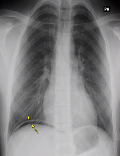
You can see fluid in the minor fissure on the right side. Blunting of costophrenic angles.
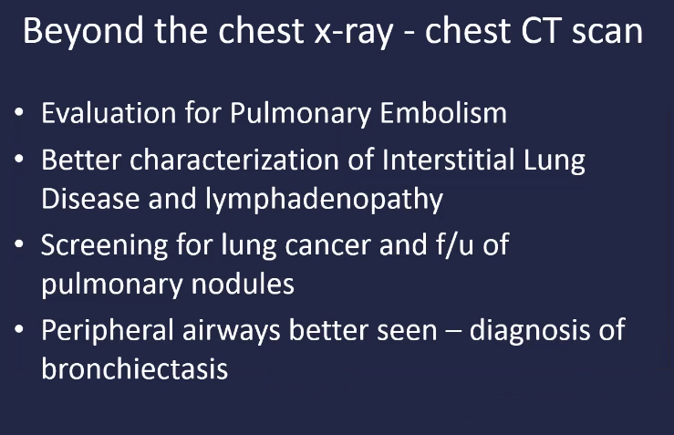
There are plenty of times when a CT scan is needed. From personal clinical experience. Always start with a cheaper and easier to obtain chest x-ray and you will be amazed what you can find with clinical context.

Post by Roger D. Struble Jr. MD MPH